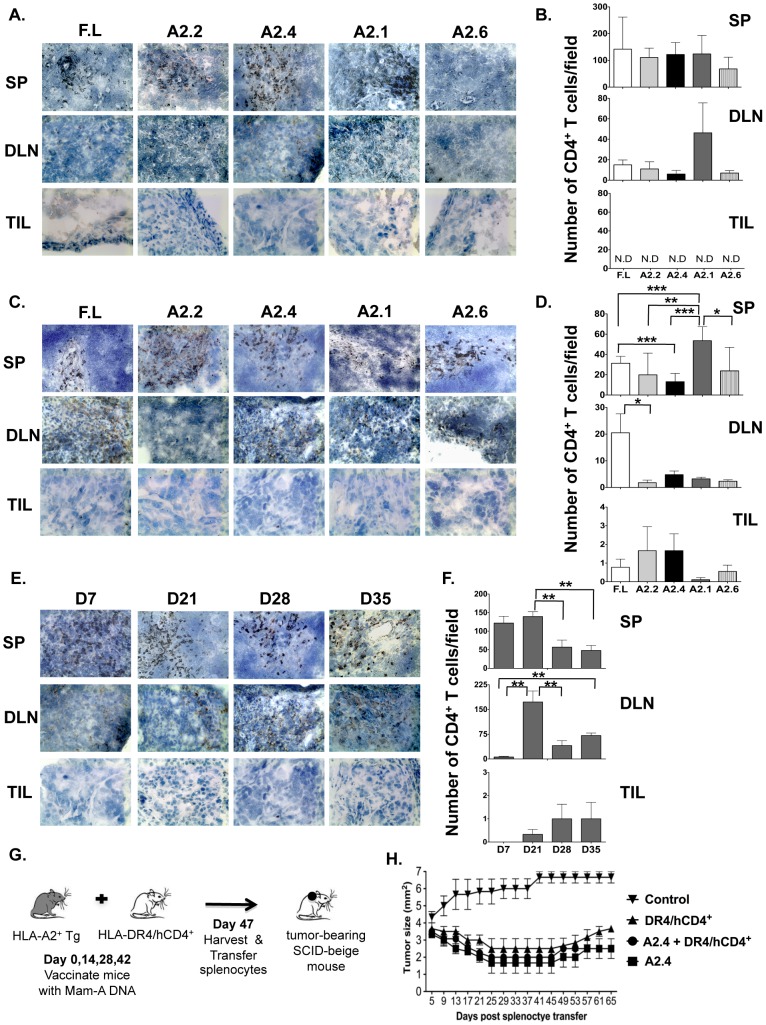
Figure 5. CD4 T cells fail to infiltrate the tumor by day 7.
SP, DLN and TIL sections from SCID-beige mice receiving splenocytes from HLA-A2+ transgenic mice vaccinated with cDNA encoding either full-length or Mam-A epitopes were analyzed by immunohistochemistry for the presence of CD4 T cells (A) 7 days and (C) 28 days post transfer (magnification of 20×). Morphometric analysis was also performed (B) 7 and (D) 28 days post transfer and results are expressed as the mean (±SEM) of 3 different experiments. ND = none detected (E–F) SP, DLN and TIL tissue sections from tumor inoculated SCID-beige mice that received splenocytes from HLA-A2+ transgenic mice vaccinated with cDNA encoding Mam-A2.4 were analyzed for the presence of CD4 T cells on days 7, 21, 28 and 35 post spleen cell transfer. (G–H) HLA-DR4+/hCD4+ transgenic mice and HLA-A2 transgenic mice were vaccinated i.m a total of 4 times, separated by 2 week intervals, with 100 µg Mam-A full-length or Mam-A2.4 specific cDNA (respectively). Five days after the last vaccination spleen cells were harvested from full-length cDNA vaccinated HLA-DR4+/hCD4+ transgenic mice and Mam-A2.4 cDNA vaccinated HLA-A2+ transgenic mice. 1×10⁷ spleen cells from either Mam-A2.4 vaccinated mice, HLA-DR4+/hCD4+ vaccinated mice or 1×10⁷ spleen cells from both Mam-A2.4 and HLA-DR4+/hCD4+ vaccinated were injected i.p into SCID-beige mice bearing human breast tumors derived from the AU-565 breast cancer cell line. Tumor-bearing SCID-beige mice without spleen cell transfer were used as controls. Tumor regression was monitored using calipers every four days (n = 4 mice/group). Data are representative of 2 experiments. * P<0.05, ** P <0.01 and *** P<0.0001.

