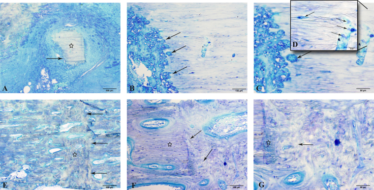
Fig. 5.
Representative photomicrographs of calcified histologic sections, comparing fracture-healing at three months after osteotomy in the control and vancomycin cohorts at ×2 (Figs. 5-A and 5-E), ×10 (Figs. 5-B and 5-F), ×20 (Figs. 5-C and 5-G), and ×40 (Fig. 5-D) magnification. The controls (Figs. 5-A through 5-D) had evidence of bone sequestrum formation (Fig. 5-A, star) with inflammatory aggregates of necrotic neutrophils resulting in cortical lysis (Figs. 5-A and 5-B, arrows) at the level of the osteotomy and multiple colonies of bacterial clusters (Figs. 5-C and 5-D, arrows), consistent with a delayed union. In comparison, vancomycin-treated plates had uniform woven bone formation (Fig. 5-E, star) across the osteotomy gap (Fig. 5-E, arrows), with formation of bridging callus consisting of both lamellar bone (Figs. 5-F and 5-G, star) and woven bone (Figs. 5-F and 5-G, arrows). Further evidence of normal bone resorption and haversian remodeling is present at the interface between the cortex and the osteotomy gap, consistent with normal fracture-healing (Fig. 5-G, arrow and star).

