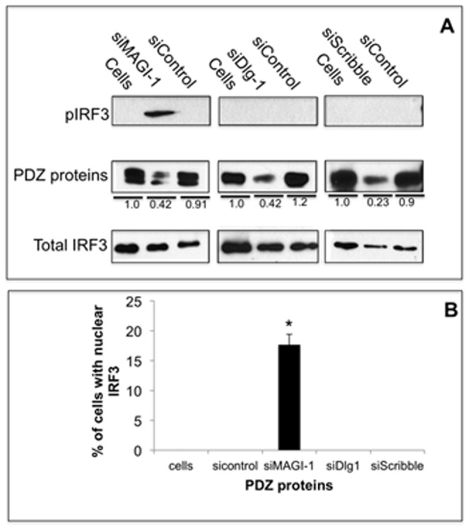
Figure 2. Depletion of MAGI-1 activates IRF3.

A) A549 cells were transfected with the indicated siRNAs against MAGI-1, Dlg-1, Scribble, and control siRNAs; lanes labeled “Cells” are non-transfected control cells. After 48 hours, cells extracted were prepared and the levels of indicated proteins were examined in immunoblots. The levels of proteins were measured by densitometry analysis using ImageJ software; the signal for each PDZ protein in non-transfected control cells was arbitrarily assigned a value of 1.0 and PDZ protein levels in transfected cells is shown relative to this 1.0 value. B) A549 cells were transfected with the indicated siRNAs. After 24 hours, cells were processed for immunofluorescence for total IRF3 as described in Methods for three independent experiments. Percentages of cells with nuclear localized-IRF3 were quantified by manually counting of at least 200 cells (a representative image for this assay is shown in Figure 7C). Error bars represent the standard error of the mean from three independent experiments, with each experiment containing duplicate samples.
