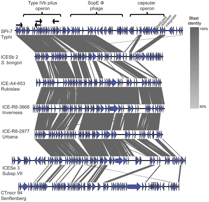
Figure 7. Comparison, made using the Blast algorithm, of SPI-7 in S. Typhi, ICES1 identified in this study, and previously sequenced ICE.
At the top is SPI-7 of S. Typhi with its three main regions (i.e., type IVb pilus operon, SopEΦ phage and capsular operon). Black arrows indicate the position of the primers used to validate the presence of the type IVb pilus operon in ICES1 in S. Rubislaw, S. Inverness and S. Urbana. Blue arrows are the coding regions and grey shaded are regions that present homology.

