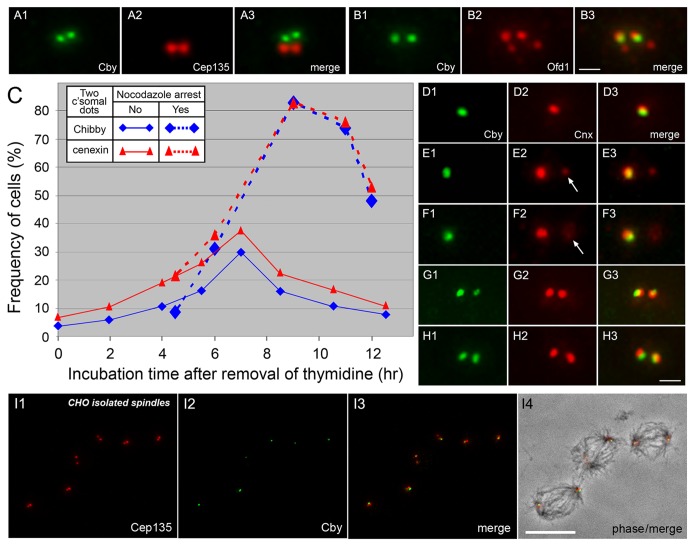
Figure 5. Emergence of Cby at the second site of the centrosome during the cell cycle. A, B:
CHO cells double stained with Cby and Cep135 (A) or Ofd1 (B) antibodies show the presence of two Cby-containing centrioles. C–H: Time course analysis of Cby/Cnx emergence in partially synchronized CHO cells arrested at G1/S or S phase by thymidine treatment. Two Cby (blue) or Cnx (red) dots were counted after removal of thymidine at time zero (C). Dotted lines indicate cells treated with nocodazole that was added at 4.25 h after washing out thymidine. D–H show double staining of centrioles with Cby and Cnx antibodies in cells fixed at 0 (D), 6 (E–F), and 7 (G–H) hr after removal of thymidine. Arrows indicate the position of Cnx-specific centrioles. I shows isolated CHO spindles stained with Cep135 and Cby antibodies and a merged image with phase-contrast is shown in I4. Cby is seen at one of the two centrioles at each spindle pole. Bars, 1 µm (A–B, D–H) and 10 µm (I).

