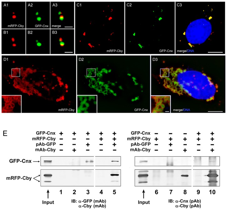
Figure 7. Interaction of Cby with Cnx.
A–D: U2OS cells expressing mRFP-Cby and GFP-Cnx at the centriole (A–B) and cytoplasmic foci (C–D). Insets of D show a high magnification of the outlined area. Bars, 1 µm (A–B, insets of D) and 10 µm (C–D). E: Co-immunoprecipitation of mRFP-Cby and GFP-Cnx expressed in HEK293T cells. Proteins were pulled down with polyclonal GFP or monoclonal Cby antibodies and blotted with monoclonal GFP/Cby and polyclonal Cnx/Cby antibodies.

