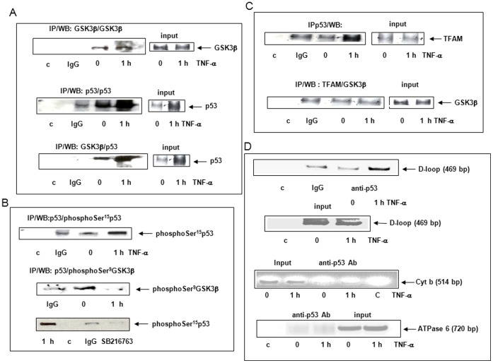
Figure 5. TNF-α induced p53 interaction to GSK3β, TFAM and D-loop.
Cells were treated for 0 (zero-time control) or 1 h with 30 ng/ml TNF-α. Mitochondrial fractions were isolated and co-immunoprecipitated or not (input) with GSK3β, p53 (FL-393) or TFAM polyclonal antibodies, IgG or no antibody was used as a control (c). (A) Western Blots were performed using GSK3β or p53 (DO1) antibody. (B) Western Blots were performed using phosphoSer15p53 or phosphoSer9GSK3β antibody in not pretreated or pretreated cells with GSK3β inhibitor SB216763. (C) Western Blots were performed using TFAM or GSK3β antibody. (D) The mtDIP assay was performed with cross-linked DNA prepared from treated cells for 0 (zero-time control) or 1 h with 30 ng/ml TNF-α. Immunoprecipitates were performed without (control, c) or with IgG used as a control or with p53 antibody. PCR were realized on immunoprecipitates or inputs using a primer pair covering D-loop, cytochrome b (cyt b) or ATPase 6 (Figure 5D).

