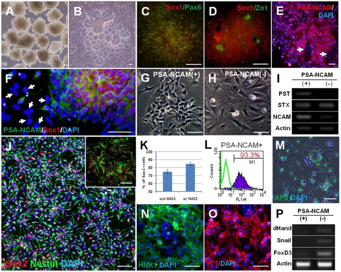
Figure 1. Efficient induction of neural rosette cells and isolation of hNPCPSA-NCAM+.
(A) hESC derived-EBs treated with dorsomorphin (DM) and SB431542 (SB), (B) When EBs treated with DM and SB were attached onto the Matrigel-coated dish, a large number of rosette structures formed in the center of the colonies after 4–5 days. (C–D) Typical neural markers such as Sox1 and Pax6 were strongly expressed in neural rosettes (C) with strong expression of Zo-1 in the central lumens of rosettes (D). (E–F) PSA-NCAM was highly expressed on the surface of neural rosette cells, but not on the cells out-migrating from the rosette clump (indicated with white arrows in E and F). (G–H) PSA-NCAM-positive cells showed typical morphology of NPCs (G), whereas PSA-NCAM-negative cells had flat and large cell bodies, similar to neural crest cells (H). (I) NCAM and PST, a polysialylating enzyme, were more abundantly expressed in PSA-NCAM-positive fraction after cell sorting. (J) A representative image for the culture of hNPCPSA-NCAM+ . After cell sorting, most of the cells were Sox2/Nestin double-positive, indicating a highly homogeneous culture of NPCs (J, inset) hNPCPSA-NCAM+ spontaneously formed the neural rosette structure. (K–L) After sorting, the proportion of Sox1-positive cells and PSA-NCAM-positive cells were enriched up to ∼85% and ∼93% of the total cells, respectively. (M-P) Cells in PSA-NCAM negative-fraction were predominantly positive for AP2 (M), HNK1 (N), and P75 (O) which were used to identify neural crest cells, and enhanced the expression of several genes implicated in neural crest development, such as dHand, Snail, and FoxD3 (P). Scale bars: 50 µm.

