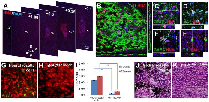
Figure 6. Integration and survival of hNPCPSA-NCAM+ in adult rat striatum.
(A) A series of coronal sections from +1.08 to −0.1 shows survival and integration of hNPCPSA-NCAM+ grafts cells in the host striatum 4 weeks after transplantation. (B) Dual-label confocal immunofluorescence microscopy with DAPI confirms a large population of HNA- and Tuj1-positive cells within the graft of hNPCPSA-NCAM+. (C–D) In addition to Tuj1-positive cells, GFAP- and NG2-positive cells within HNA-positive graft sites show the potential of hNPCPSA-NCAM+ in differentiating into all three neural lineages in vivo. (E–F) Further dual immunohistochemistry for HNA:GABA and HNA:TH indicates the ability of grafted cells to differentiate into various neuronal subtypes. (G–H) A representative area of a neural rosette cell graft indicates a significantly higher number of Ki67-positive cells and clusters whereas only a few Ki67-positive cells with no clusters are observed for hNPCPSA-NCAM+. (I) Bars indicate the proportions of Ki67-positive cells in the grafts of neural rosette cells and hNPCPSA-NCAM+ at 4 weeks (blue bars) and 12 weeks (red bars) post-transplantation as mean ± s.e.m. (J–K) Representative images of H/E-stained brain tissues grafted with unsorted neural rosette cells (J) and hNPCPSA-NCAM+ (K). Melanocyte-like cells stained in dark-brown color were shown only in the grafts of unsorted neural rosette cells (J). Scale bar: 50 µm. Abbreviations: LV, lateral ventricle; A, anterior; P, posterior; D, dorsal; V, ventral. *, p<0.05.

