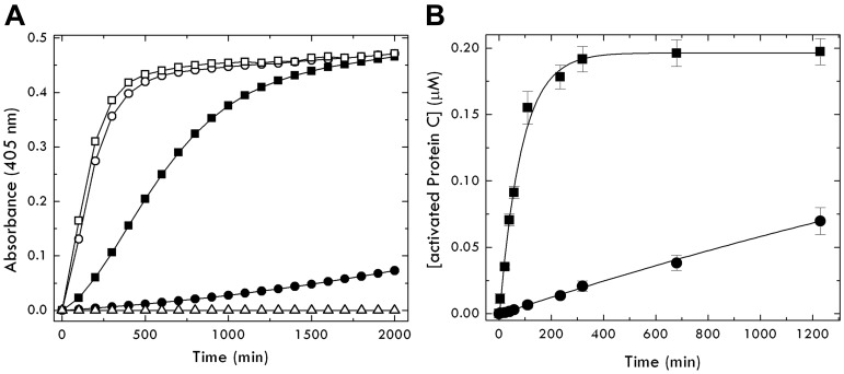
Figure 2.
Activation of the EDD mutant of protein C by thrombin. (A) Shown are progress curves of DRR hydrolysis by activated protein C generated from the zymogen form (circles represent wild-type, and squares, EDD) on interaction with thrombin, in the absence (closed symbols) or presence (open symbols) of 50nM thrombomodulin, under experimental conditions of 5mM Tris, pH 7.4, 145mM NaCl, 5mM CaCl2, 0.1% PEG8000 at 37°C. Analysis of the curves gives the value of kcat/Km for activation of protein C as follows: 0.11 ± 0.01mM−1s−1 (●), 6.9 ± 0.1mM−1s−1 (■), 190 ± 10mM−1s−1 (○), and 560 ± 20mM−1s−1 (□). ▵ represents the inactive EDDS mutant of protein C as a control. The sigmoidal nature of the progress curve, most visible in the case of the EDD mutant, is the result of the continuous nature of the assay32 that measures hydrolysis of DRR after the buildup of activated protein C. Autoactivation of EDD is negligible under the conditions used in the assay because it evolves over a time scale that extends beyond the complete generation of activated protein C by thrombin (Figure 1). (B) Kinetics of protein C wild-type (0.2μM, ●) and mutant EDD (0.2μM, ■) activation by thrombin (30nM) under conditions of pseudo–first order kinetics ([protein C] ≪ Km). Continuous lines were drawn according to a single exponential with values of kobs/[thrombin] = kcat/Km of 0.11 ± 0.02 (wild-type) and 6.7 ± 0.3 (EDD mutant), in excellent agreement with the values determined independently from progress curves of DRR hydrolysis (see A). No lag phase is observed in this discontinuous assay31 because activation of protein C is determined by quenching the thrombin-catalyzed reaction at different times under pseudo–first order kinetics.

