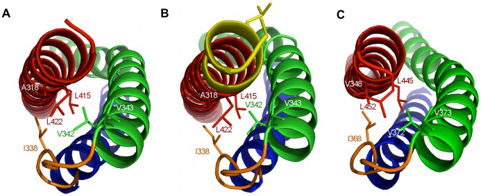
Figure 3. Interactions formed by the C-terminus of utrophin and dystrophin spectrin repeat one domains.
Structural representations of A) Utr-SR1, B) Utr-SR1-L and C) Dys-SR1 showing the burial of hydrophobic sidechains (stick representation) on the B helix (green) and A–B loop (orange) by the C-terminus of helix C (red). The A helix is coloured blue. The extended Utr-SR1-L C-terminus and sidechains of the SR2 heptad repeat (L427, L430) are coloured yellow. The protein main-chain is depicted in ribbon representation with key side-chains shown as sticks.

