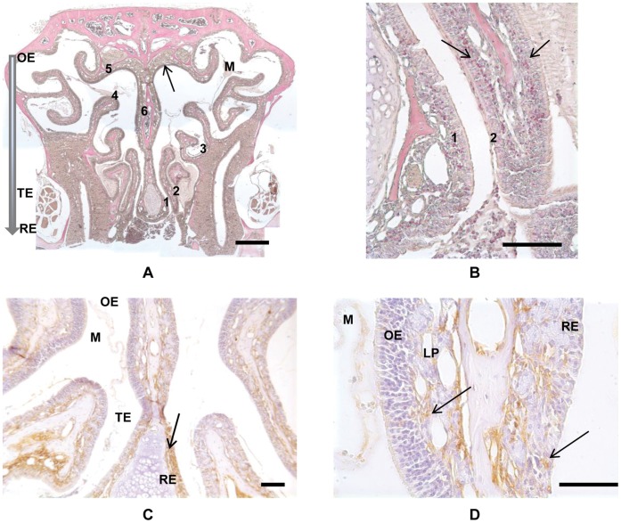
Figure 1. Demonstration of eosinophils and major basic protein (MBP) in a chronic allergen exposed mouse.
A) Low power image of Sirius Red and hematoxylin stained coronal section of a chronic allergen exposed mouse. Numerals identify the six sampling areas, where specific 100 micron square areas were designated for eosinophil counting. The solid arrow indicates the region of the olfactory cleft where ion concentration measurements were made, in the dorsal recess near sampling area 5. The shaded arrow indicates the general dorsal to ventral transition from Olfactory (OE), Transitional (TE) to Respiratory (RE) epithelia in the mouse nose. Mucus is evident within the lumen (M). Bar = 500 µm. B) Higher power image from panel 1 showing counting regions 1 and 2. Arrows indicate Sirius Red stained eosinophils localized to Lamina Propria (LP) and Respiratory Epithelium (RE). Bar = 100 µm. C) Localization of MBP in the coronal section of a chronic allergen exposed mouse. Primary antibody is mouse anti-MBP and non-specific binding is blocked by mouse IgG incubation. Brown staining indicates MPB (arrow). Regions of Olfactory (OE), Transitional (TE) and Respiratory (RE) epithelia are indicated. Mucus is evident within the lumen (M). Bar = 50 µm. D) Higher power image from panel 3. Arrows indicate MBP localized to Lamina Propria (LP) and Respiratory Epithelium (RE). Olfactory epithelium (OE) is largely devoid of MBP. Mucus (M) does not show any significant non-specific staining. Bar = 50 µm.

