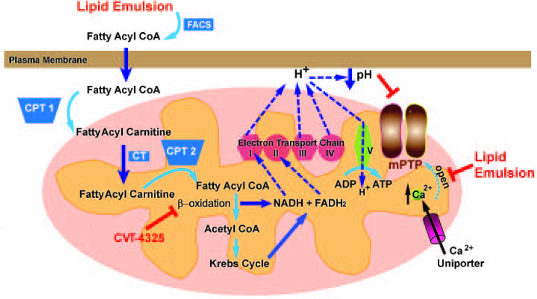
Figure 5. Proposed mechanisms underlying LE-induced rescue of cardiac arrest as a result of bupivacaine-overdose.
Oxidation of LE contains long chain fatty acids occurs in the mitochondria. Fatty acids are transported into the mitochondria via carnitine shuttle, and go through β-oxidation to produce NADH and FADH2. NADH and FADH2 enter the electron transport chain (ETC). ETC couples electron transfer between an electron donor (NADH and FADH2) and an electron acceptor (such as O2) to the transfer of H+ ions (protons) across a membrane. The resulting electrochemical proton gradient is used to generate energy in the form of adenosine triphosphate (ATP) in complex V. The release of H+ from complex I, III and IV lead to lowering PH in outer mitochondrial membrane which lead to inhibition of mPTP opening. Inhibition of β-oxidation pathway by CVT abolishes the production of NADH, FADH2, as well as H+ ions, leading to generation of less ATP. The rise of Ca2+ ions in mitochondria through Ca2+ uniporter will trigger the opening of mPTP. LE enhances the homeostasis of cardiomyocytes to better regulate calcium overload and therefore delay the opening of mPTP. FACS; Fatty Acyl CoA Synthase, CPT1, Carnitine Acyl Transferase 1, CT; Carnitine Acylcarnitine Translocase, CPT2, Carnitine Acyl Transferase 2.

