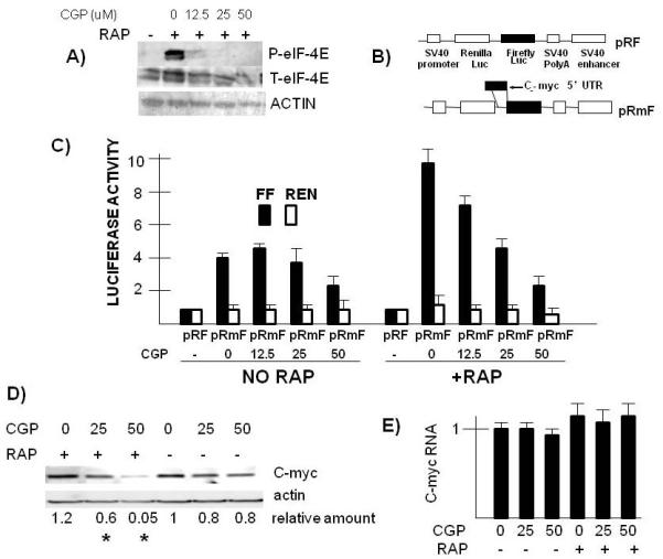
Figure 3.

Effect of MNK inhibitor on myc IRES activity and myc expression. A) ANBL-6 MM cells pre-treated with the CGP MNK inhibitor for 30 mins at varying concentrations. Followed by addition of rapamycin (100 nM) for 3 hrs and then immunoblot assay for phospho-eIF-4E, total eIF-4E and actin. B) Reporter constructs used to assay for myc IRES activity. C) Firefly (FF, dark bars) or Renilla (Ren, open bars) luciferase expression in pRF versus pRmF-transfected ANBL-6 cells treated+/−rapamycin (100 nM) and +/− CGP (used at 0, 12.5, 25 or 50 uM). All lluciferase activity is normalized to the luciferase values (both Renilla and Firefly) obtained for pRF in the absence of added rapamycin and CGP (designated “1”). Results represent means+/−SD of quadruplicate samples. D) ANBL-6 MM cells pre-treated with CGP at 0, 25 or 50 uM followed by addition of rapamycin (or not) at 100 nM for 8 hrs. Immunoblot assay then performed for c-myc or actin expression. Relative amount of c-myc protein determined densitometrically (ratio of c-myc/actin) and represents means of three independent experiments. Asterix denotes a value significantly lower (p<0.05) than control (no CGP). E) Experiment performed as in “D” but assay is real time PCR for c-myc RNA expression (data are means+/−SDs of 3 experiments).
