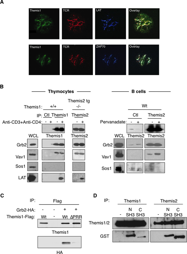
Figure 5.

Themis1 and Themis2 are recruited within Grb2/Vav1 signaling complexes to LAT following TCR engagement. A, Formation of TCR activation clusters in Jurkat T cells expressing Themis1-GFP protein plated onto anti-CD3 coated coverslips. The colocalization of Themis1-GFP (green) with the TCR (red), ZAP70 (blue) and LAT (blue) were analyzed by confocal microscopy. B, Analysis of LAT, Vav1, Sos1 and Grb2 co-immunoprecipitation with Themis1 and Themis2 in thymocytes and B cells. Thymocytes (Themis1+/+ or Themis1−/−; Themis2tg) or B cells were stimulated with anti-CD3+anti-CD4 antibodies or pervanadate respectively. Anti-Themis1 (thymocytes) or anti-Themis2 (thymocytes and B cells) immunoprecipitates were analyzed by western blot with the indicated antibodies. C, The proline rich region of Themis1 mediates its association with Grb2. 293T cells were transfected with expression plasmids encoding Themis1-Flag, Grb2-HA, or Themis1-ΔPRR (deleted of the RxPXXP motif) in the combinations shown. Anti-Themis1 immunoprecipitates were blotted with anti-HA and anti-Themis1 antibodies. D, Themis1 and Themis2 interact with the N-terminal and the C-terminal SH3 domains of Grb2. Thymocyte lysates from Wt or Themis2 transgenic mice were incubated with GST fusion protein expressing the N-terminal SH3 domain (1–68) or the C-terminal SH3 domain (156–199) of Grb2. Themis1 or Themis2 were immunoprecipitated and co-precipitation of GST-fusion proteins were analyzed by immunoblot with the indicated antibodies. Results shown are representative of three experiments.
