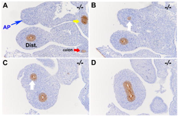
Figure 5. E-cadherin positive cells are not detected within the duodenal atretic precursor at E12.0 (CS17).

E-cadherin staining of the duodenal atretic precursor of an Fgfr2IIIb-/- embryo at E12.0 (A-D) and in a stage matched wild-type embryo (E-H). Images are organized in a rostral to caudal fashion. A. E-cadherin positive cells are absent in the atretic precursor (AP) region (blue arrow) and present in the region distal to this (B and C, white arrow) and in the C- loop of the duodenum (D). In contrast E-cadherin positive cells are seen lining a continuous luminal structure throughout the duodenum proximal and distal to the C-loop in the wild-type control (E-H). (Bars indicate 100μM). Yellow arrows indicate pancreatic duct tissue. Red arrow indicates colon. Pink arrow indicates pylorus. Dist. Indicates distal duodenum beyond the C-loop.
