
FIG. 3.
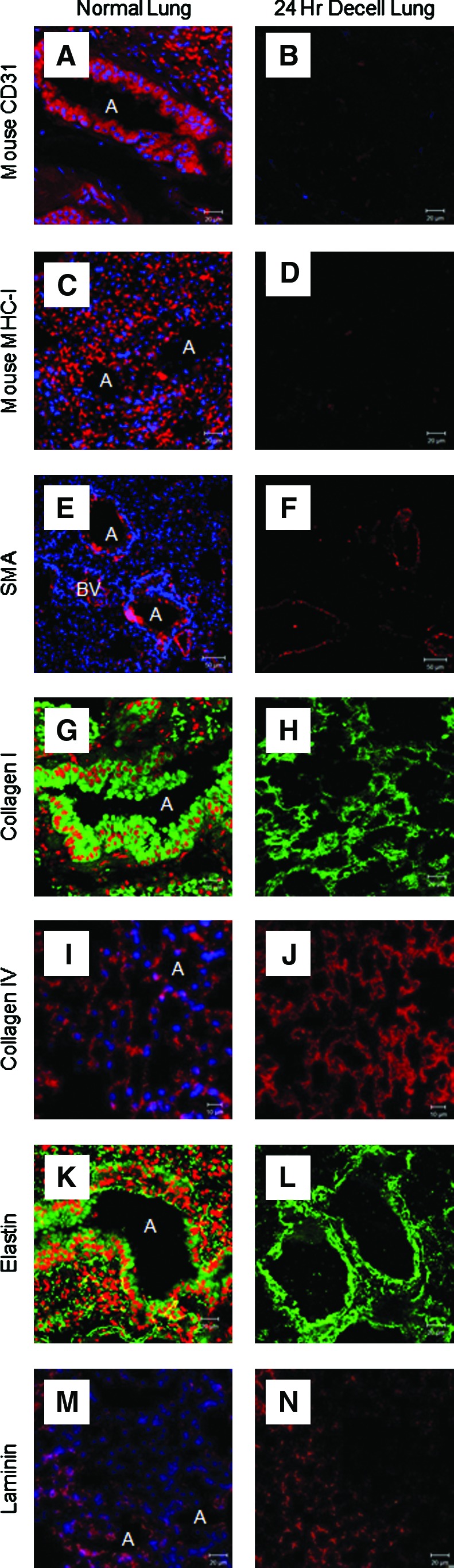
An immunofluoresence analysis of a de-cellularized scaffold following the 24-h de-cellularization protocol shows preservation of ECM proteins with loss of endothelial cell and MHC class I expression. Mouse CD31 (A, B), mouse MHC Class I (C, D), SMA (E, F), collagen I (G, H), collagen IV (I, J), elastin (K, L), and laminin (M, N). Representative images for all conditions are shown (63×oil magnification). ECM, extracellular matrix; SMA, smooth muscle actin; A, airway; BV, blood vessel. Color images available online at www.liebertonline.com/tec