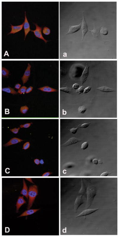
Fig. 3.

Confocal fluorescence microscopy of paxillin in PC3 cells during cell attachment. Cells were cultured in complete medium, or Tyr/Phe-, Gln-, or Met-restricted medium for 3 days and then allowed to attach to fibronectin coated four-well slides for 4 h. The slides were incubated with an anti-paxillin monoclonal antibody and then stained with a Texas Red-conjugated horse anti-mouse IgG antibody. The slides were then mounted with an anti-fade mounting medium containing DAPI to show nuclear staining. A and a: Confocal fluorescence (A) and phase contrast (a) images in PC3 cells cultured in normal medium. B and b: Cells cultured in Tyr/Phe-free medium for 3 days. C and c: Cells cultured in Met-free medium for 3 days. D and d: Cells cultured in Gln-free medium for 3 days. [Color figure can be viewed in the online issue, which is available at www.interscience.wiley.com.]
