
Figure 1.
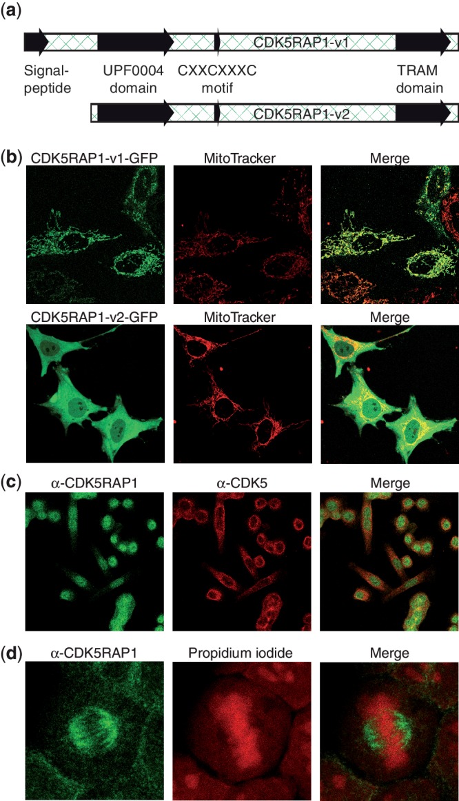
Subcelluar distribution of CDK5RAP1 variants. (a) Representation of the two investigated CDK5RAP1 variants with mitochondrial import sequence and catalytic domain. (b) Live-cell images of HeLa cells transfected with C-terminally GFP-tagged CDK5RAP1-v1 and CDK5RAP1-v2. For the counterstain of mitochondria, cells were incubated with MitoTracker Red dye. The merge shows that CDK5RAP1-v1 colocalizes with mitochondria, while CDK5RAP1-v2 is distributed in both cytoplasm and nucleus. (c) Formaldehyde-fixed HeLa cells were stained with an antibody against the N-terminal region of CDK5RAP1-v2 (Alexa-488, green) and against CDK5 (Alexa-555, red). The localization of CDK5RAP1 is predominantly nuclear with little overlap to CDK5. (d) Again, fixed HeLa cells were stained with anti-CDK5RAP1 (green). For localization of nucleic acids, propidium iodide (red) was used. CDK5RAP1 shows an association with the mitotic spindle.