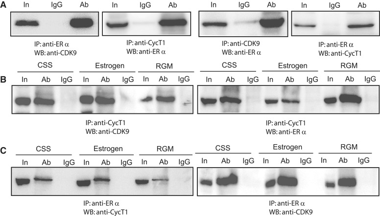
Figure 4.
Complex formation between CyclinT1, CDK9 and ERα. (A) Expression plasmids encoding CyclinT1, CDK9 and ERα were co-transfected into HEK293 cells and immunoprecipitated (IP) with the indicated primary antibodies and the resultant complexes were analyzed by western blotting (WB) using the indicated secondary antibodies. (B) and (C) Identification of complex formation by endogenous proteins. MCF-7 cells were grown in estrogen free (CSS), 10 nM estrogen added and in regular growth medium (control). Extracts, prepared from cells grown in the three different conditions, were used for immunoprecipitation (IP) with anti-CyclinT1 in (B) and anti-ERα in (C). Complexes were analyzed by WB using the antibodies indicated under each panel. In each case, 10% of the total cell extract was loaded for comparison and is labeled ‘In’ (input).

