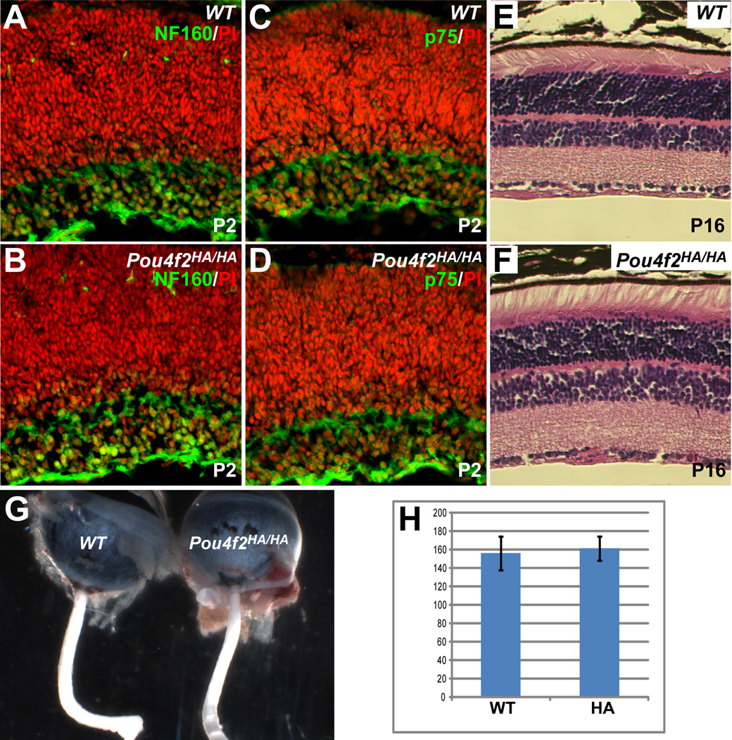
Figure 5.
RGCs develop normally in Pou4f2HA/HA mice. (A–D) Staining for two RGC markers, NF160 and p75 (green), at P2 revealed that there was no obvious change in the numbers of RGCs or expression levels of the two proteins in Pou4f2HA/HA retinas. Red is nuclear staining by PI. (E, F) H&E staining shows that mature (P16) Pou4f2HA/HA retinas have normal structures as compared to wild-type (WT); the cell numbers in all three layers are comparable with those of wild-type controls. (G) The optic nerves of Pou4f2HA/HA mice are not reduced in size compared to the wild-type. (H) No significant difference is seen in the total numbers of cells between the GCLs of Pou4f2HA/HA and wild-type mice at P2 (n=4, p=0.34). Y axis is number of cells per arbitrary length unit.

