Figure 1.
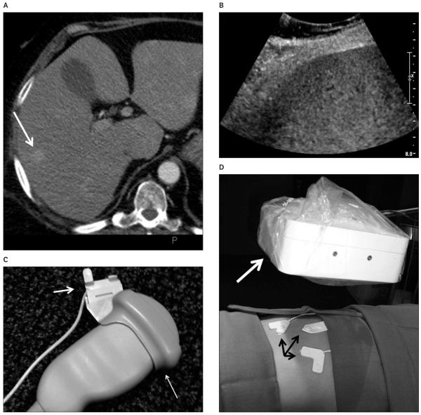
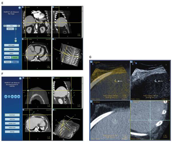
Corticotropin-producing neuroendocrine tumor in a 61-year-old woman. A, Arterial phase computed tomography shows an enhancing liver lesion (arrow). B, Sonography of the liver shows no discernable mass. C, Ultrasound transducer with a disposable electromagnetic tracking sensor (short arrow) attached to a sterilizable needle guide (long arrow). The sterile cover can go above or below the sensor. D, The electromagnetic field generator (white arrow) is mounted on an articulated arm and covered with a sterile drape. Fiducial patches or markers (black arrows) on the skin provide references for registration. E, The graphical user interface shows the lesion in 3 planes, which may be helpful for confirming selection of the center of the target in all planes. F, Skin fiducials (markers) are identified and confirmed to ensure accurate registration, ie, matching of anatomic points on the skin coordinates within the imaging data. G, The tracking screen during needle placement displays the relationship between the targeted lesion (T) and the needle tip (arrow).


