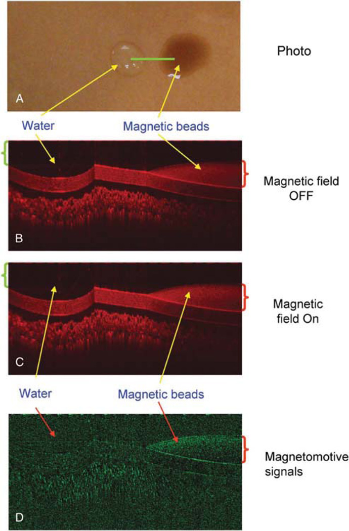
FIG. 2.
Optical coherence tomography (OCT) imaging of water and magnetic particles. One drop of water (green bracket ~50 µL) was instilled onto a plastic card surface along with another drop (red bracket ~ 50 µL) of the magnetic particles. The card was placed on a magnetic coil. An OCT linear scan (A, green line) was performed. The OCT imaging was repeated during the magnetic modulation OFF (B) and ON (C). Magnetomotive signal in M-model (D) showed only the magnetic particles with a great reduction of the background.

