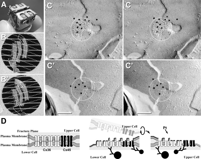
Fig. 3.
Explanation of the DR-FRIL technique. a Photograph of the DR stage after fracturing of two specimens. Matched DR samples (indicated by B and B′ in a) are shown at higher magnification in b, b′), after floating off into buffer. b, b′ Replicated but undigested samples were mounted on thin-bar grids, with matching outlines indicated. Small tissue fragments were lost during washing (open outlines opposite corresponding outlined tissues). Bars occluding matching areas are indicated by dotted lines. c, c′ One of 11 pairs of matched gap junction hemiplaques (circles with inscribed quadrants) from the samples indicated in b, b′. Cx45 is labeled with 5-nm gold beads (lower right quadrants), whereas Cx36 is labeled with 10-nm gold beads (upper left three quadrants c, c′). Labeling for Cx45 is aligned with (i.e., opposite) labeling for Cx45 in the matching areas of the two hemiplaques. Likewise, labeling for Cx36 is aligned with labeling for Cx36. These matching hemiplaques demonstrate bihomotypic gap junctions. b, c Modified from Li et al. (2008a). d Diagram showing production of matched DRs and the subsequent immunogold labeling of bihomotypic plaques containing mostly Cx36 (white connexons labeled with large gold beads) and fewer Cx45 (black connexons, labeled with small gold beads)

