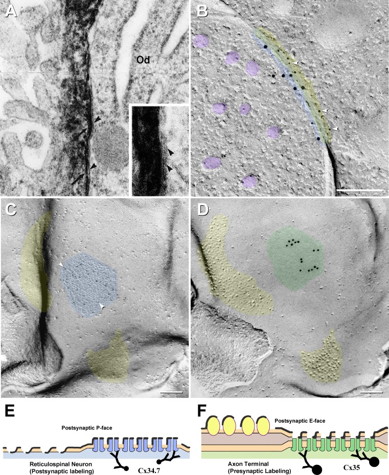
Fig. 4.
Comparison of heterotypic labeling in TEM thin sections (a), cross-fractured FRIL images (b) and by the DR-FRIL technique (c, d), with explanatory drawing (e, f). a Thin-section immunocytochemical demonstration of Cx43 in the astrocyte side of an O:A gap junction, labeled by the peroxidase–antiperoxidase method, leaving DAB deposition on the astrocyte side (arrows) and the oligodendrocyte side (Od) unlabeled (arrowheads). [This image was obtained 10 years before Cx47 was identified as the coupling partner for Cx43; modified from Ochalski et al. (1997)]. b Cross-fractured “mixed” (electrical plus chemical synapse), presumably from an auditory afferent onto an unidentified reticulospinal neuron in goldfish hindbrain. In this companion image to (c), 5-nm gold beads labeled postsynaptic connexin Cx34.7 (arrowheads), whereas 10-nm gold beads for Cx35 labeled presynaptic connexins. (Synaptic vesicles are indicated by purple overlays.) The yellow overlay indicates the radius of uncertainty of immunogold labeling for small gold beads, the blue overlay indicates the radius of uncertainty for large gold beads and the green overlay indicates the region of potential overlap. This asymmetric distribution of gold labels reveals that this gap junction between a sensory afferent and the reticulospinal neuron is heterotypic. c, d Matching complementary replicas at club ending synapse on reticulospinal neuron. The postsynaptic hemiplaque (c, designated by blue overlay) is labeled for Cx34.7 by approximately three 5-nm gold beads (arrowheads), whereas the complementary E-face (green overlay) is labeled for Cx35 by 15 10-nm gold beads. Areas corresponding to glutamate receptor–containing postsynaptic densities are indicated by yellow overlays, with the P-face pits in c matching the E-face particles in (b), as previously shown by labeling for glutamate receptors (Pereda et al. 2003). e, f Diagram of matching replica complements, showing Cx34.7 without Cx35 in the reticulospinal neuron (e) and Cx35 labeling without labeling for Cx34.7 in the E-face of the matching hemiplaque of the apposed club ending. Labeling in (d) and (f) is for connexins in the cytoplasm of the underlying axon terminal ending, even though only the E-face pits of the reticulospinal neuron are visualized in the replica

