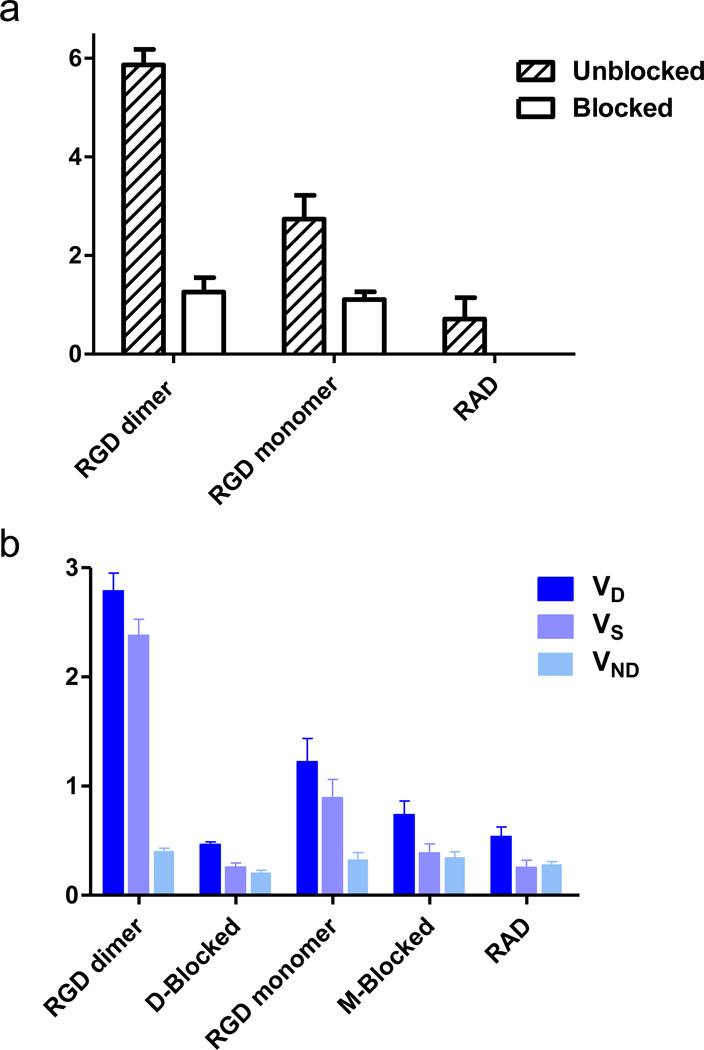
Fig. 5.
a Binding potential (BpND) of 18F-labeled RGD peptide tracers. b volumes of distribution (VT) of 18F-labeled RGD peptide tracers. The binding potential was calculated as BpND = k3/k4 reflecting the binding affinity, and the volume of distribution (VT = K1/k2(1+ k3/k4)) reflects the tissue-to-plasma concentration ratio. VT can be regarded as the sum of specific (VS = K1·k3/(k2·k4)) and nonspecific (VND = K1/k2) distribution.

