Figure 3.

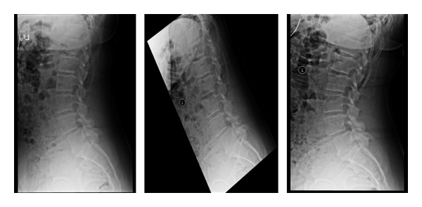
Lateral X-rays demonstrating a L4-L5 grade 1 spondylolisthesis. The standing lateral dynamic flexion and extension X-rays are used to determine motion at this segment.

Lateral X-rays demonstrating a L4-L5 grade 1 spondylolisthesis. The standing lateral dynamic flexion and extension X-rays are used to determine motion at this segment.