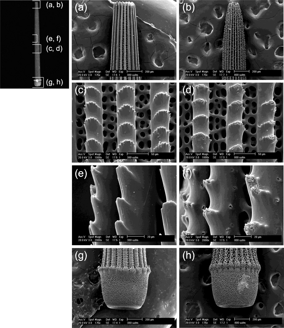
Figure 2.
Scanning electron micrographs of Lytechinus variegatus spines from individuals reared at ambient (380 µatm) and elevated (800 µatm) pCO2 over the course of 89 days. The regions of the spines shown are indicated on the representative spine at left. Scale bars for each view are indicated on SEM micrographs. (a,b) lateral view of spine tip, showing the control (a) in contrast to thinning of longitudinal striations at 800 µatm (b); (c,d) middle of spine (front view) comparing the fenestrated structure and longitudinal striations of the control (c) with evidence of malformation and/or dissolution at 800 µatm (d); (e,f) middle of spine (high magnification, side view), showing normal skeleton (e) and evidence of malformation and/or dissolution at 800 µatm (f); (g,h) base of spine and calcareous annular ridge in the control (g) compared to thinning of longitudinal striations at 800 µatm (h).

