
Figure 2.
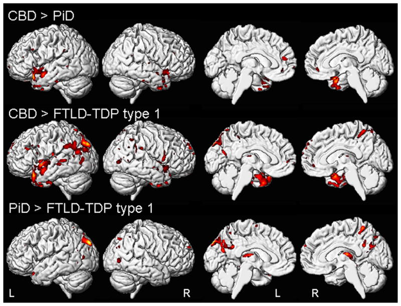
Regions of grey matter loss that were different between the bvFTD subjects with PiD, CBD or FTLD-TDP type 1 pathology. The top panel shows regions of greater loss in PiD compared to CBD; the middle panel shows regions of greater loss in FTLD-TDP type 1 compared to CBD; and the bottom panel shows regions of greater loss in FTLD-TDP type 1 compared to PiD. Results are shown uncorrected for multiple comparisons at p<0.001 on medial and lateral three dimensional renderings of the brain.