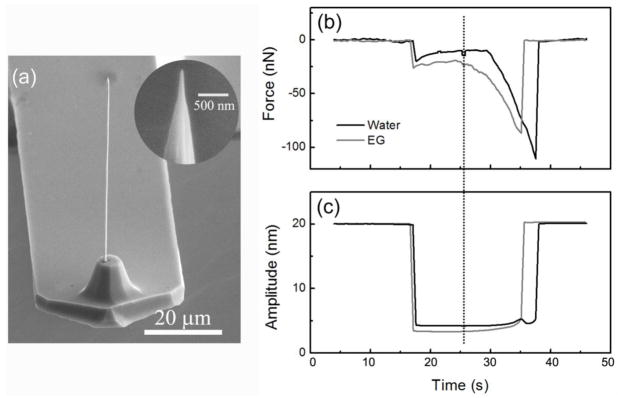
Fig. 2.
(a) Image showing a ~60 μm long and ~600 nm-diameter Pt nanowire directly deposited onto an AFM cantilever with the meniscus-confined electrodeposition technique. The inset shows the tip end of the needle sharpened down to ~25 nm in tip radius of curvature with focused ion beam milling. (b–c) Static approach and retraction curves of the needle tip into water and ethylene glycol showing the change of the exerted force due to surface tension measured from the cantilever deflection (b) and the change of the cantilever oscillation amplitudes (c). The dashed line marks the starting point of the retraction in the experiment.

