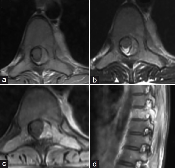
Figure 1.

Axial T1-weighted (a) and T2-weighted (b) magnetic resonance imaging at D9 level showing the lesion involving the left half of the vertebral body, pedicle, transverse process, and the lamina with an epidural component producing cord compression. Postgadolinium injection axial and sagittal T1-weighted images (c and d) show intense enhancement of the tumor. Note the enhancing component in the paraspinal thoracic region
