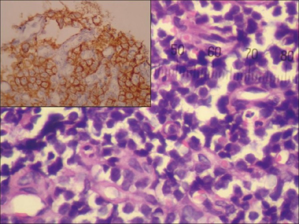
Figure 3.

Microphotograph showing a highly cellular tumor, consisting mainly of small round to oval cells with hyperchromatic nuclei and remarkably scanty cytoplasm along with the presence of Homer-Wright pseudorosettes; the tumor cells being immunopositive for CD 99 (inset)
