
Fig. 4.
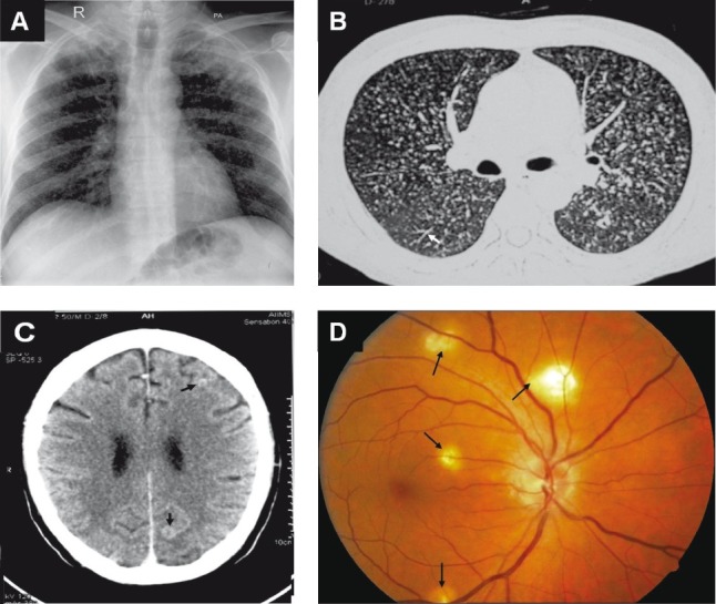
Chest radiograph (postero-anterior view) (A) and chest CT (lung window) (B) showing classical miliary pattern, tree-in-bud appearance (B) (arrow). The patient also had cerebral tuberculomas (arrows) and TB meningitis (C). Choroid tubercles, located in the posterior pole of the orbit (D) (arrows) offered an early valuable clue to the diagnosis. The present case illustrates the importance of documenting the presence of neurological involvement, particularly, TB meningitis in patients with miliary TB and thereby ensuring adequate duration of anti-tuberculosis treatment and need for corticosteroid treatment.