
Fig. 9.
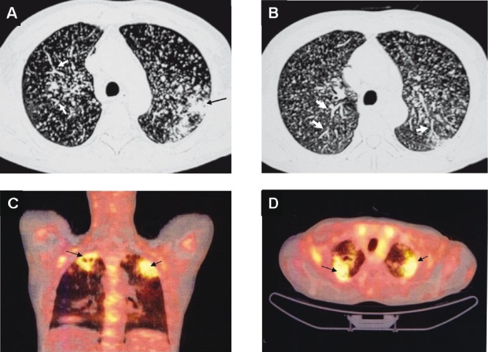
Chest CT (lung window) of the same patient as in Fig. 4 showing pulmonary parenchymal lesions (black arrow) (A). In addition to the miliary pattern, well-defined, linear, branching opacities (tree-in-bud appearance) (thick white arrows) (A and B) are also seen. This pattern is evident when centrilobular bronchioles are dilated, or, are filled with mucus, fluid or, pus and represents endobronchial spreading of infection. 18FDG-PET CT of the same patient showing increased activity in the pulmonary parenchymal lesions (arrows) but not in the miliary lesions (C and D). The 18FDG-PET has potential to further understanding the clinico-radiographic-functional correlation in miliary tuberculosis and merits further study. However, it may not be useful in intracranial TB. CT, computed tomography; 18FDG-PET CT, 18F labelled 2-deoxy-D-glucose positron emission tomography-computed tomography; TB, tuberculosis.