
Figure 2.
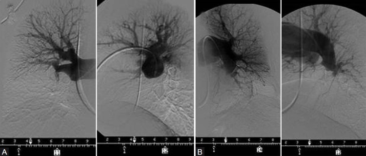
(A) PA and lateral right pulmonary angiogram of the patient whose V/Q scan is shown in Figure 1; complete obstruction of the right interlobar vessel. (B) PA and lateral left pulmonary angiogram, showing a “pouch” occlusion of the descending pulmonary vessel beyond the superior segment; appreciated on the lateral view is a small lingular artery which is difficult to discern A B on AP view.