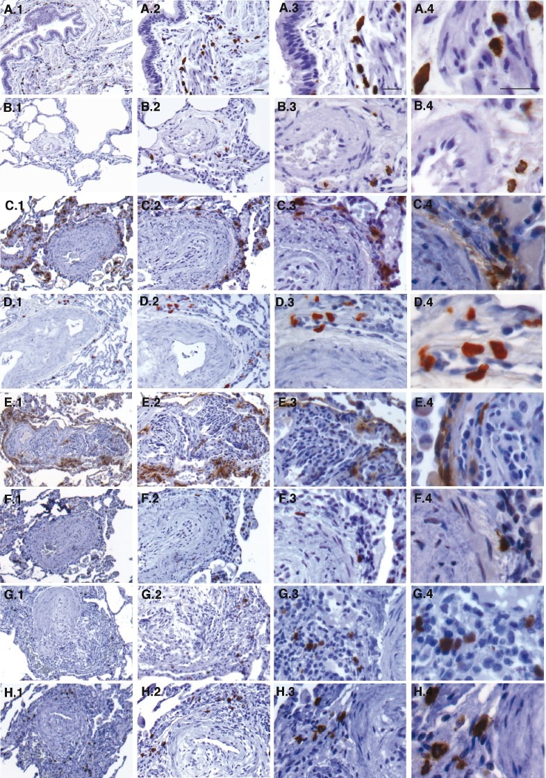
Figure 1.

Increased numbers of connective tissue type mast cells in PAH lungs. (A–H) Tryptase and chymase immunostaining of explanted control and PAH lungs. (A–E) Tryptase stained lung tissue sections. (F–H) Chymase stained lung tissue sections. Explanted PAH and failed donor (control) lung paraffin embedded tissue sections were immunostained for tryptase and chymase to identify and characterize mast cell phenotype (brown cells). Mast cell numbers were increased in the lungs of patients with PAH compared to controls (P=0.01) and localized predominantly to perivascular regions as opposed to submucosal regions as in control lungs. Mast cells in PAH lungs were tryptase+ and chymase+ consistent with a connective tissue phenotype as opposed to primarily tryptase+ in control lungs. Panels A–B – Control lungs stained for tryptase: (A) Mast cells are seen in the submucosal regions of the airways of control lungs. (B) Mast cells around a blood vessel in a control lung are less compared to PAH lungs. Panels C–H – PAH lungs: (C–E) Tryptase+ mast cells in PAH are in the perivascular adventitia and increased in number. (F–H) Mast cells in the perivascular regions are chymase+. In panels E–G where plexiform lesions are noted, mast cells are seen within the lesions. Magnification: (1) 5×, (2) 10×, (3) 20×, (4) 40×. Scale bar: 25μm.
