Figure 1.

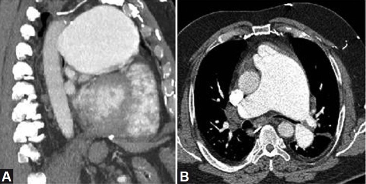
CT images showing massive dilatation of the proximal pulmonary artery on sagittal (A) and axial (B) images, with severe compression of the left mainstem bronchus.

CT images showing massive dilatation of the proximal pulmonary artery on sagittal (A) and axial (B) images, with severe compression of the left mainstem bronchus.