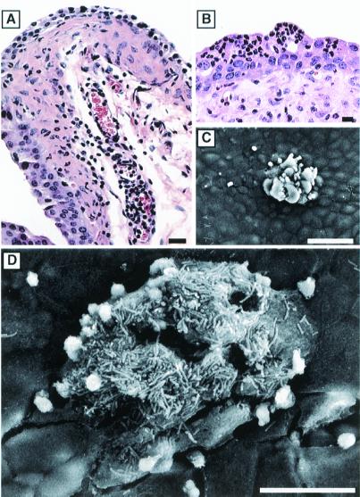
Figure 6.
Neutrophil influx into the urothelium in response to infection. (A) Paraffin sections of C57BL/6 mouse bladders recovered 6 h after infection with type 1-piliated UPEC and stained with hematoxylin and eosin show PMNs (small, darkly stained cells) migrating from blood vessels within the lamina propria and into the urothelium. (B) PMNs appeared to aggregate beneath the luminal surface of the bladder and could occasionally be seen, by scanning EM, emerging in the vicinity of adherent bacteria on the surface of newly exposed immature urothelial cells (C). PMNs were also found associated with infected facet cells in the process of exfoliating (D). [Bars = 20 μm (A), 10 μm (B), 50 μm (C), and 30 μm (D).]

