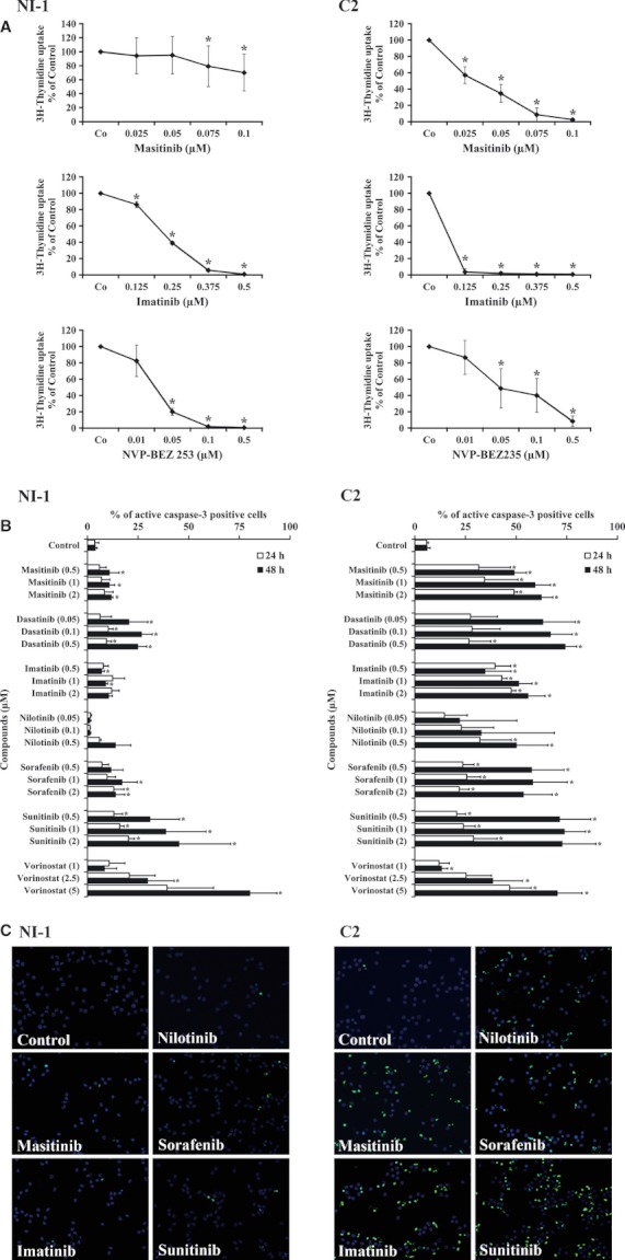
Effects of various drugs on proliferation and apoptosis of NI-1 cells and C2 cells. (A) NI-1 cells (left panels) and C2 cells (right panels) were incubated in control medium or with various concentrations of targeted drugs (masitinib, imatinib, NVP-BEZ235, as indicated) at 37°C for 48 h. Then, 3H-thymidine uptake was measured as described in the text. Results show the percent of control and represent the mean ± SD of at least three independent experiments. Asterisk: P < 0.05. (B) NI-1 cells and C2 cells were incubated in control medium or in medium containing various concentrations of targeted drugs (as indicated) at 37°C for 24 h (open bars) or 48 h (black bars). Then, cells were recovered, and the expression of activated caspase-3 was assessed by flow cytometry. Results show the percentage of active caspase-3-positive cells and represent the mean ± SD of three independent experiments. Asterisk: P < 0.05. (C) TUNEL assay experiments using NI-1 cells, C2 cells, and various targeted drugs. Cells were incubated in control medium or in medium containing masitinib (2 μM), imatinib (2 μM), nilotinib (2 μM), sorafenib (2 μM), and sunitinib (2 μM) as indicated (37°C, 24 h). The TUNEL assay was performed as described in the text. As visible, the targeted drugs applied induced apoptosis in C2 cells but not in NI-1 cells, confirming results obtained by flow cytometry with an antibody against active caspase-3.
