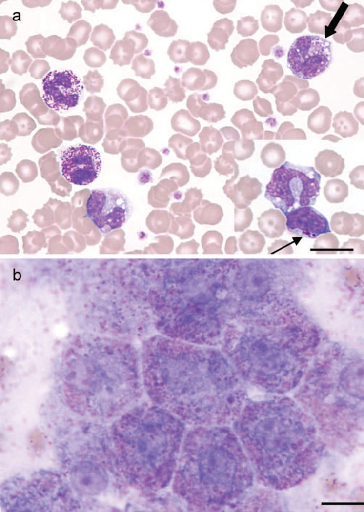
Figure 2.
(a) Blood smear from a Miniature Poodle-like dog diagnosed with mucopolysaccharidosis type VI showing metachromatic Alder-Reilly-type inclusions in neutrophils and macrophage (thick arrow), with similar inclusions in lymphocytes (inset, thin arrow) (Leishman stain, bar = 12 µm). (b) Impression smear of liver from the same dog taken at time of necropsy, showing pink-purple inclusions in hepatocytes (Leishman stain, bar = 10 µm).

