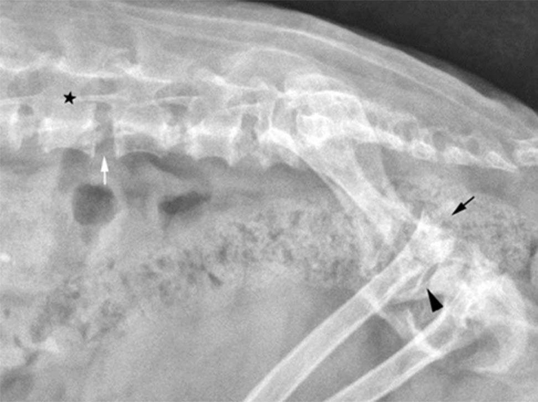
Figure 3.
Lateral radiograph of the lumbosacral spine and pelvis of a Miniature Poodle-like dog, diagnosed with mucopolysaccharidosis type VI showing a reduction in length of the vertebral bodies with incomplete ossification of the end plates. The intervertebral disc spaces (white arrow) and vertebral canal (star) appear widened. There is severe subluxation of the sacrum with respect to L7 and dorsal luxation of one of the femoral heads (black arrow). The contralateral coxofemoral joint space is markedly widened (arrowhead).

