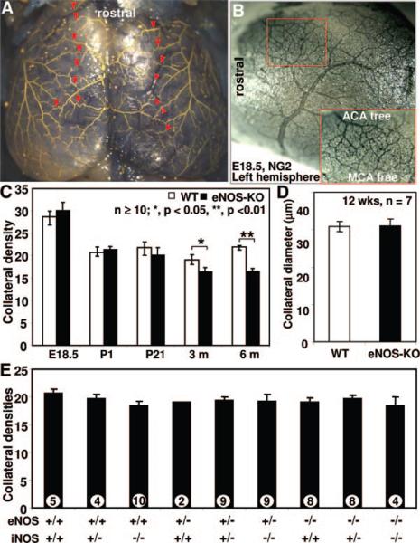
Figure 5. Cerebral cortical pial collateral number and diameter at different developmental stages and different genotypes.
A, Representative image of MicrofilP-casted adult eNOS-KO mice showing pial artery network and collaterals. Red arrowheads indicate collaterals between middle cerebral (MCA) and anterior cerebral artery (ACA) branches counted and diameters determined. B, representative image of E18.5 eNOS-KO mouse embryonic brain. C and D, Collateral number and diameter. E, No significant differences in pial collateral number in P28 pups crossed to harboring indicated alleles. *P<0.05, **P<0.01; 2-tailed t test.

