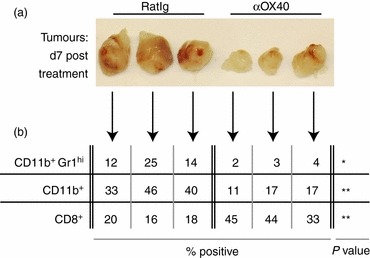
Figure 1.

MCA205 cells were injected subcutaneousl in C57BL/6 mice and allowed to grow to 5–7 mm in diameter (approximately day 10). Mice were then injected intraperitoneally with 250 μg αOX40 or control RatIg and the tumour was removed 7 days later. (a) Photographs of individual isolated tumours are shown. (b) The percentage CD11b+Gr1hi IAlo immature macrophages, the overall CD11b+ macrophage population, and the percentage infiltrating CD8+ T cells from those same tumours are provided. NS: not significant; *P < 0·05; **P < 0·01; ***P < 0·001.
