
Figure 3.
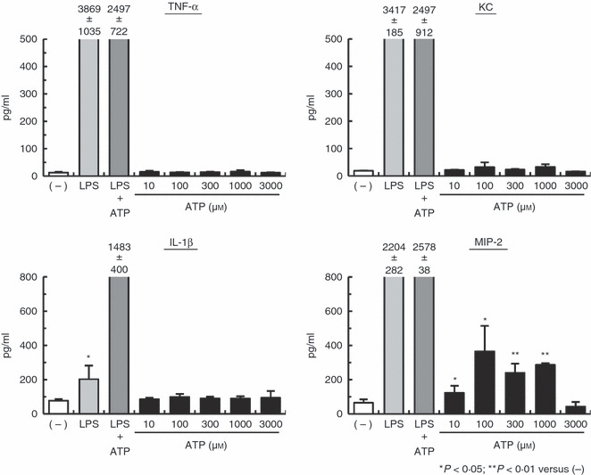
ATP stimulation of macrophages in vitro leads to macrophage inflammatory protein-2 (MIP-2) production. Peritoneal exudate macrophages (PEMs) were primed with 1 μg/ml lipopolysaccharide (LPS) for 4 hr and then stimulated with or without ATP for 24 hr. Culture supernatants were harvested and assayed for interleukin-1β (IL-1β), tumour necrosis factor-α (TNF-α), keratinocyte-derived chemokine (KC) and MIP-2 by ELISA. Data represent the mean ± SEM of triplicate samples. Experiments were repeated three times.