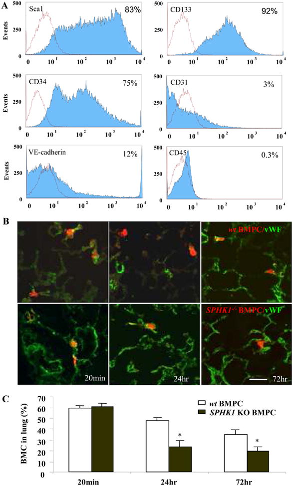
Figure 1. FACS analysis and lung uptake of mouse bone marrow-derived progenitor cells (BMPCs).

A) FACS analysis was performed using cultured BMPCs on day#21 for hematopoietic progenitor/stem cell markers (Sca-1, CD133, and CD34), endothelial cell markers (CD31, and VE-cadherin), and myeloid markers (CD45). Histogram plots for these markers are shown in blue and negative control antibody results are shown in red. Results are representative for 3 experiments. B) Morphological assessment of BMPC sequestration in lungs. Rhodamine-labeled BMPCs were localized within lung microvessels as evident by BMPCs surrounded by lung vascular endothelial cells stained with FITC-conjugated von Willebrand Factor (vWF) at 20 min post-injection (left panel) and in lung parenchyma distinct from vessel lumen at 24 hr and 72 hr after injection (right panel, bar=30μm) C) Quantification of total number of CMTMR-labeled wt BMPCs and SPHK1-/-BMPCs at 20min, 24hr and 72hr following cell injection.
