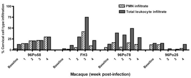
Fig. 1.
Relative increase in inflammatory cells in cervical cell collections of sexually transmitted infection (STI)-infected macaques. Analyses were conducted in Chlamydia trachomatis-only (96Po58)- and C. trachomatis + Trichomonas vaginalis–infected macaques (FH3 and 96Po78). 96Po25 received mock media inoculations (control). Cell populations were collected by cervical cytobrush sampling and enumerated by microscopy utilizing an Endtz-trypan differential stain. The graph longitudinally depicts the percent of cervical infiltrate cell types present at baseline and weeks 1–4 post-infection (week 1, relative to first C. trachomatis inoculum). An increase in the percentage of inflammatory cells consisting of polymorphonuclear (PMN) cells and/or total leukocyte infiltrate, relative to baseline, was observed in all three sexually transmitted infection (STI)-infected macaques over the course of the study.

