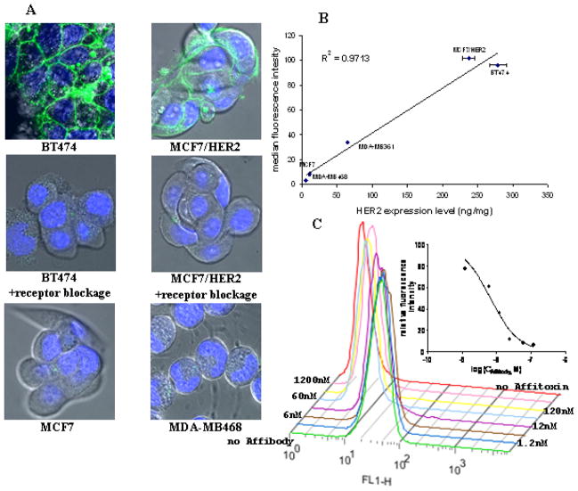
Figure 4. Specificity of Affitoxin binding confirmed by confocal imaging and flow cytometry.
A. Cells were incubated with 0.1 μM of Alexa Fluor® 488-labeled Affitoxin for 6 hours with and without Affibody molecules. B. HER2 expression level in five different breast cancer cell lines was determined by ELISA and is expressed in nanograms of HER2 per milligram of protein lysate. Cells were incubated with 5 μg/ml of Alexa Fluor® 488-labeled Affitoxin. Binding efficacy was determined by flow cytometry. C. BT474 cells were incubated with 5 μg/ml of Alexa Fluor® 488-labeled Affitoxin and increasing concentrations of Affibody. Binding efficacy was determined by flow cytometry and quantified by GraphPad software.

