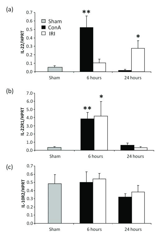
Figure 1.
IL-22 signaling in ConA and IRI models in WT mice at 6 h and 24 h of reperfusion. Quantitative RT-PCR-assisted analysis of (a) IL-22; (b) IL-22R1; and (c) IL-10R2 expression in mouse liver tissue subjected to ConA (N=6/group; 15 μg/g i.v.) or IR-triggered damage (90 min ischemia: N=6/group; Sham N=4/group). (Statistical comparisons between experimental and sham samples *: p<0.05, **: p<0.005). (a) Mice subjected to ConA injection expressed high levels of IL-22 at 6 h of infusion, before returning to baseline at 24 h, while IRI mice had low levels of IL-22 at 6 h and a modest increase by 24 h. (b) IL-22R1 expression was elevated in both models at 6 h, and returned to baseline at 24 h. (c) IL-10R2 was not affected by ConA or IRI.

