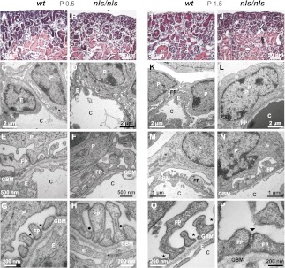
Figure 3.
Destruction of the glomerular filtration barrier in nls/nls mice. (A, B, I and J) Hematoxylin and eosin–stained paraffin sections of renal cortex from (A and I) wt and (B and J) nls/nls mice. Whereas morphologic changes are not obvious at P0.5 in nls/nls mice (B), renal corpuscles with enlarged urinary space (white arrow) and dilated tubules with cast formation in the lumen (black arrow) are evident at P1.5 (J). (C, D, K, and L) Low-power TEM of renal corpuscles from (C) wt and (D) homozygous mutant at P0.5 and from (K) wt and (L) nls/nls mice at P1.5 showing the glomerular filtration barrier. No differences are obvious in immature podocytes at P0.5 in nls/nls mice (D), but dramatically effaced FPs are observed at P1.5 (L). (E–H and M–P) TEM of glomerular filtration barrier of wt (E and G) and homozygous mutants (F and H) at P0.5 showing regular slit membranes (asterisk) and FPs in wt (G), but either immature or malformed FPs with apically located slit-like junctions (black dots) in mutants (H). At P1.5, FPs with mature slit diaphragms (asterisk) are evident in wt (M and O) but immature FPs with occluding junctions or ladder-like structures (arrowhead) are observed in nls/nls mice (N and P). TEM, transmission electron microscopy; C, capillary; E, endothelial cell; P, podocyte.

