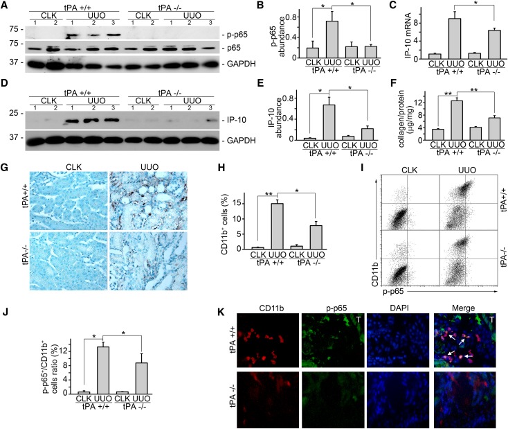
Figure 6.
tPA mediates NF-κB activation in vivo. tPA knockout mice displayed significantly reduced induction of (A and B) phospho-p65 and (C) interferon-inducible protein-10 mRNA and (D and E) protein production. (A and D) Representative Western blots. Number indicates individual mouse. (B, C, and E) Quantitative analysis of relative abundance. *P<0.05 (n=5 mice per group). (F) Total renal collagen content was evaluated by Sirius Red/Fast Green collagen assay kit and expressed as micrograms collage per milligram protein. **P<0.01 (n=5 mice per group). (G) Representative immunohistochemical staining of CD11b for macrophages. (H) Quantitative demonstration of interstitial infiltration of CD11b-positive macrophages. *P<0.05, **P<0.01 (n=5 mice per group). (I) Representative flow cytometry analysis. Upper right indicates CD11b-positive macrophages with activated NF-κB signaling. (J) Quantitative analysis of relative amount of CD11b macrophages with activated p65 phosphorylation. Unit was expressed as percentage of both phospho-p65– and CD11b-positive cells in gated cells. *P<0.05 (n=4 mice per group). (K) Double immunostaining of phospho-p65 (green) and CD11b (red) in the diseased kidney after 7 days obstruction. Nuclei were indicated by DAPI. Original magnification, ×400. Cells with both positive staining of phospho-p65 and CD11b were indicated by arrows. Tubular staining was marked with T. CLK, contralateral kidney.

